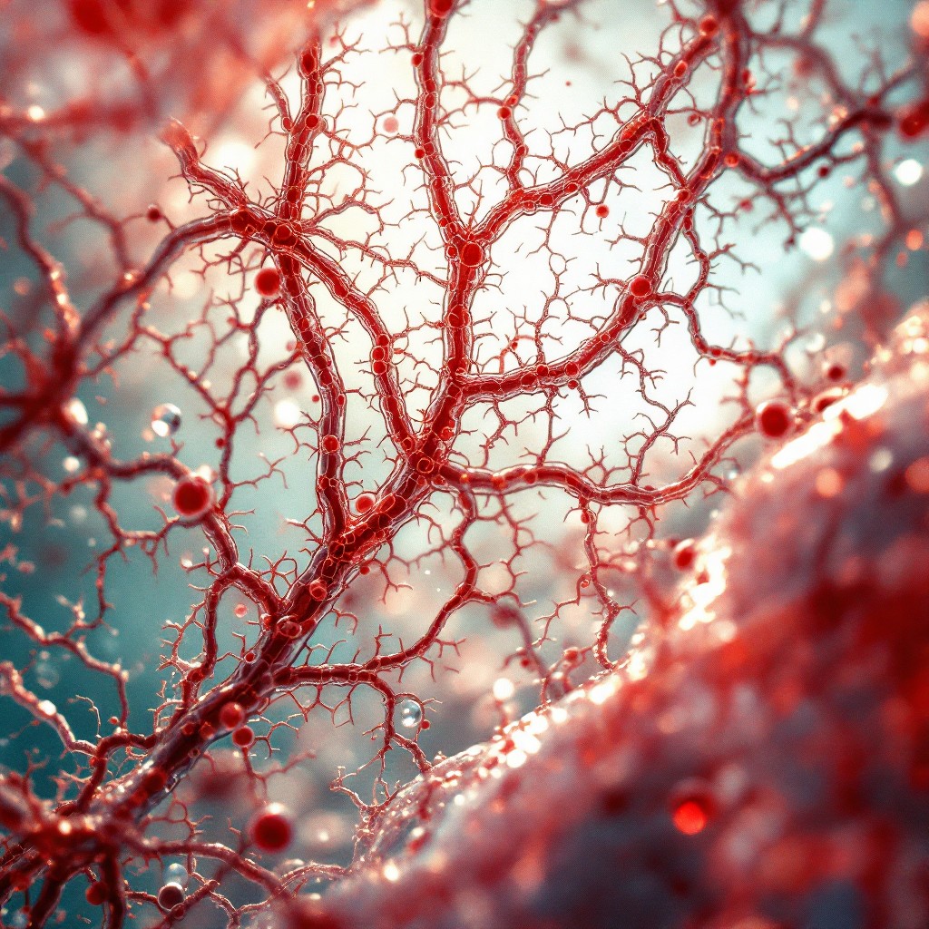
To investigate potential treatments for different diseases, researchers usually start by using either animal models or human cell cultures. However, animal models do not always mimic human diseases well, and cell cultures fall far short of the complexity of real tissue. Advances in 3D printing, combined with knowledge of biomaterials, are enabling scientists to recreate complex 3D tissue models in the laboratory.
The Hybrid Biofunctional Materials group at CIC biomaGUNE, led by Ikerbasque Researcher Dorleta Jimenez de Aberasturi, is studying how to combine advanced bioprinting techniques with nanomaterials to obtain tissue models containing vessels with different layers that can respond when external stimuli are applied.
The body’s tissues are irrigated by blood vessels that provide them with oxygen and nutrients, which makes it essential to develop new methods to fabricate them. The more realistic organ and tissue models are, the better researchers can understand the causes of different diseases, test the efficacy of new therapeutic compounds, or even use them as grafts.
Drs. Dorleta Jimenez de Aberasturi, Uxue Aizarna, and Malou Henriksen from CIC biomaGUNE have made important advances in the field of 3D bioprinting to develop materials that allow the fabrication of tissue models increasingly close to real tissues for laboratory research.
To fabricate these materials, high-precision printers are used, and their “ink” must meet specific requirements. One of the challenges in this field is to find materials suitable for printing that also have optimal properties for cell survival. Both the printing method and the properties of these materials are crucial to producing high-quality tissues quickly and accurately, which is why this field of research continues to innovate in pursuit of better results. It is also important, as Jimenez de Aberasturi notes, to “find materials that can be combined to enable multimaterial printing and thus create stable structures with different layers of each material.”
Improved materials for more complex structures
In collaboration with a group at Maastricht University (The Netherlands), the CIC biomaGUNE team has achieved, through embedded bioprinting, the ability to “print soft or liquid materials that cannot be printed in air, as they would deform or collapse before solidifying. The ink used in this type of bioprinting favors the survival of the embedded cells,” explains Jimenez de Aberasturi. Using this technique, they have fabricated a blood vessel model with concentric cylinders that mimic the different layers of an artery.
In addition, to develop a model artery with valves, the research team collaborated with a group at Utrecht University (The Netherlands) using volumetric bioprinting, which “consists of forming the entire volume of the structure at once by projecting design images into a specific volume of resin or developed material, followed by curing (hardening) of the material, instead of building it layer by layer. This technique makes it possible to achieve complex shapes at high speed,” adds the CIC biomaGUNE researcher. The researchers were able to design and precisely print “a valve that can open and close when an external stimulus is applied, similar to those in the heart.” As a result, “we could eventually print a valve, an artery, or a vein by replicating the image of a real patient’s vessel.”
In both projects, methacrylated gelatin was used as a base. This is a soft material derived from gelatin that is chemically modified to become solid when exposed to light. This type of gelatin is highly useful for bioprinting, as it is cell-compatible and its viscosity can be modified. Extracellular matrix (the microscopic scaffolding found between cells that supports body tissues), obtained from porcine pulmonary artery, was added to this gelatin.
“One important but difficult aspect to recreate is the type of mechanical forces to which blood vessels are subjected due to changes in blood pressure. These changes are relevant if we want realistic cellular responses,” explains Dr. Jimenez de Aberasturi, principal investigator of the laboratory. To address this, the researchers used gold nanoparticles, which have a unique capacity to interact with light in a very intense and controllable way.
With this innovative combination of materials and bioprinting techniques, the CIC biomaGUNE team has advanced the fabrication of artificial arteries that mimic arterial pulse in response to a laser. “By combining these improved materials with different printing techniques, we have managed to fabricate more complex structures that are much more similar to those found in the human body,” they affirm. Nevertheless, they acknowledge that “this is a very complex process. Although we are now very close to making these arteries highly realistic, there are still steps to take. And each step is a world in itself.”
Bibliographic references
Hybrid Plasmonic Bioresins and dECM-Based Materials for Volumetric Bioprinting of Vascular-Inspired Architectures. Uxue Aizarna-Lopetegui, Gabriel Größbacher, Ada Herrero-Ruiz, Aitor Tejo-Otero, Malou Henriksen-Lacey, Riccardo Levato, and Dorleta Jimenez de Aberasturi. ACS Applied Materials & Interfaces Article ASAP. DOI: 10.1021/acsami.5c03880
The Route to Artery Mimetics: Hybrid Bioinks for Embedded Bioprinting of Multimaterial Cylindrical Models. Uxue Aizarna-Lopetegui, Ada Herrero-Ruiz, Monize C. Decarli, Ane Urigoitia-Asua, Karla Chavarri-Urraca, Lorenzo Moroni, Malou Henriksen-Lacey, Dorleta Jimenez de Aberasturi. Advanced Functional Materials. DOI: 10.1002/adfm.202419072
.png)
