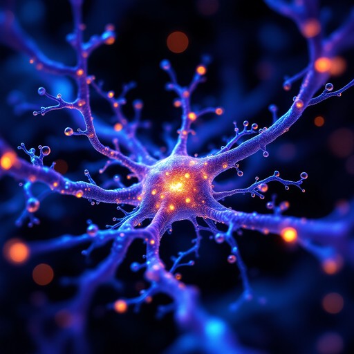
For decades, Alzheimer’s research has focused on two main protagonists: a protein fragment known as amyloid-beta, which accumulates in the space between neurons—the cells responsible for transmitting information—in people with Alzheimer’s disease; and the tau protein, which builds up inside neurons. However, there is growing evidence that these two pieces of the puzzle are not enough to fully understand the disease.
Several studies have suggested that myelin—the layer that insulates neurons and enables signals to travel rapidly between them—may be affected in the early stages of Alzheimer’s, even before symptoms become apparent.
“In recent years, there has been a shift in perspective, with increasing emphasis on the potential role of myelin degradation in the progression of the disease. These changes in myelin are believed to occur at very early stages,” explains Dr Jordi Llop, from CIC biomaGUNE. “In people with Alzheimer’s, myelin is degraded or altered in some way, although it is not yet known whether this is a cause or a consequence of the disease.”
Against this backdrop, CIC biomaGUNE’s Radiochemistry and Nuclear Imaging research group, led by Dr Llop, is collaborating with Professor Carlos Matute’s group at EHU to explore ways of repairing or preventing alterations to myelin as a strategy for treating Alzheimer’s. “The ultimate aim of our project is to investigate whether certain compounds can help restore myelin in our mouse model of Alzheimer’s and whether, over time, this leads to cognitive improvement,” adds Dr Llop.
Making the invisible visible
Changes in myelin are not easy to detect. Although brain-imaging techniques make it possible to examine the brain without surgery, they do not always capture subtle alterations in myelin, meaning that much of this degradation can remain hidden. CIC biomaGUNE has a new generation of tools that makes it possible “to observe in real time whether myelin sheaths can actually be repaired in animal models that simulate what happens over the course of the disease,” Llop explains. “We use imaging techniques to monitor in real time how the sheath surrounding neurons is protected, and we combine this with behavioural tests to determine whether the mice recover their memory or experience less memory loss.”
Positron emission tomography (PET) is an imaging technique that uses small radioactive molecules as tracers. Once inside the body, these molecules travel through the brain, making it possible to detect specific biological processes. In Alzheimer’s research, “we are using a tracer capable of highlighting areas where myelin has been damaged. When myelin deteriorates, it exposes certain structures that are normally hidden. The tracer binds to these structures and produces a detectable signal. In a way, it is as though the brain were using invisible ink to reveal the areas where something is beginning to go wrong,” explains Mariana Coimbra de Almeida, a PhD researcher in the group.
The CIC biomaGUNE team is also working to automate some of the processes used to analyse the images generated in its studies. PhD researcher Sebastián Acebal is developing artificial intelligence models: “To understand what is happening in the brain, one of the most important things is to determine what is occurring in each specific region. To do this, we divide the images obtained by my colleagues and define the regions of interest. This process, known as segmentation, is normally carried out manually, but it is extremely time-consuming and difficult. As part of my PhD project, I am developing different artificial intelligence models to automate this process, along with other programs and software aimed at improving the results,” Acebal explains.
Early detection, disease monitoring and new therapeutic strategies
Initial studies using these tools show that it is possible to detect Alzheimer’s-related changes in regions of the brain where myelin is abundant. This lends support to an increasingly prominent idea: Alzheimer’s is not only a disease involving the accumulation of proteins, but also a disorder of connectivity. Neurons do not function in isolation. They form complex networks, whose effectiveness depends on signals travelling correctly. If myelin fails, these networks begin to lose efficiency, even if the neurons themselves remain present.
The MYAMI project is seeking new treatments for Alzheimer’s disease. “A better understanding of the role of myelin opens up new possibilities,” says Llop. On the one hand, it could help detect Alzheimer’s at earlier stages, when changes are not yet clinically apparent. On the other, it could enable the progression of the disease to be monitored more accurately. It also opens the door to new therapeutic strategies. If myelin deterioration is part of the problem, protecting or repairing it could become a key treatment target.
.png)

