The brain is the most complex biological material and it evolved over hundreds of millions of years, from simple neural networks, performing simple learned behaviors, such as avoiding dangerous situations to improve the rate of survival. The essential role for memories is pivotal for survival, so it goes without saying that the constellation of interacting cells that form memory engrams must go well back along the evolutionary time scale.
However, the prevailing dogma today is that memories are formed in the hippocampus and later stored in the cortex. This limited view does not take into consideration the other brain structures, especially the evolutionarily older brain structures, performing dynamics re-organization of anatomical and functional circuits for forming and storing memories.
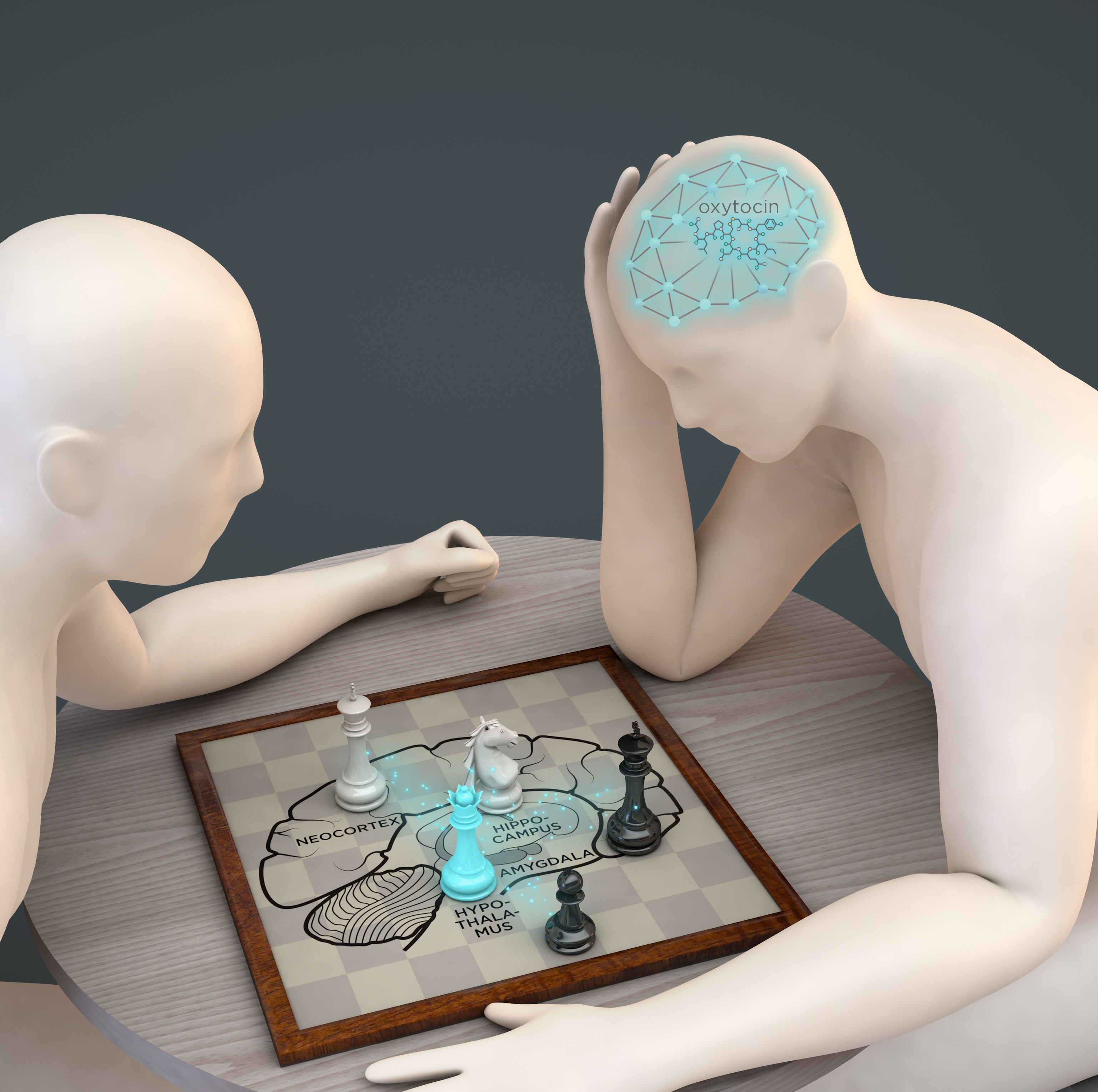
In a recent study published in the Journal Neuron, an international interdisciplinary team, lead by the Ikerbasque Researcher Mazahir T. Hasan, reasoned that memory “engram” or “trace” is likely to be formed and preserved also in the evolutionary old brain structure, such as the hypothalamus. Scientists targeted specific cell types in the hypothalamus, namely neurons producing oxytocin – a neuropeptide, that controls various emotional brain functions, including fear.
The team developed a novel genetic method to selectively tag the oxytocin neurons which are recruited during learning, memory formation and retrieval. Using this technique, the authors discovered that indeed context-specific engrams are formed and preserved in the hypothalamic circuits and perturbation of these engram circuits drastically affects fear memories.
This conclusion came from the experiments in which the authors smuggled into the hypothalamus genetic switches designed to selectively “tag” the oxytocin neurons activated during fear retrieval. These “tagged” cells were genetically loaded with viruses with engineered proteins to manipulate neuronal activity either by blue light stimulation to activate the tagged cells (called optogenetics) or by delivering a synthetic chemical to silence these neurons (called chemogenetics). When the researchers activated these tagged cells, the animals, who have learned to freeze in a dangerous environment started to move around; basically, the fear expression was blocked as long as the neurons are activated. When blue light was switched-off, the fear expression returned. This shows that the tagged cells “contain the knowledge” of fear. The authors then performed the reverse experiment by silencing the engram oxytocin neurons. They found that the same circuit is also needed to erase fear in a process called “extinction”. Remarkably, these cells undergo enormous plasticity, switching from slow transmission mediated by the neuropeptide oxytocin to fast response by the fast activating glutamate transmission.
This discovery is a “game-changer”, as it calls for action to explore the memory engrams across the different brain regions, both lower and higher brain structures. By understanding the anatomical and functional fear circuits, it should be possible to design innovative strategies to treat human mental diseases, when fear memory becomes pathological, such in the general anxiety and especially posttraumatic stress disorders.
For more information: https://www.ncbi.nlm.nih.gov/pubmed/31104950
.png)
